


Introduction
Imagine a person suddenly develops weakness on one side of the body and struggles to speak. Their family may hope the symptoms will disappear, but in stroke care, waiting can cost valuable time. Rapid recognition and treatment can make a meaningful difference to recovery.
Mechanical thrombectomy has transformed the treatment of selected patients with acute ischaemic stroke by allowing doctors to remove a blood clot blocking a major artery in the brain. Alongside advances in imaging and emergency stroke care, this procedure has expanded treatment options for patients at risk of severe disability. 1,2
What Happens When a Blood Clot Blocks the Brain?
An ischaemic stroke occurs when a blood vessel supplying the brain becomes blocked, commonly by a blood clot. Without adequate blood flow, brain cells are deprived of oxygen and nutrients and begin to sustain damage. Symptoms may include weakness, speech difficulties, visual changes or problems with balance.
A large-vessel occlusion (LVO), which blocks a major brain artery, can cause severe neurological impairment. Prompt assessment and brain imaging help clinicians identify the blockage and determine the most appropriate treatment. 1
The First 4.5 Hours: Can Medication Dissolve the Clot?
Intravenous thrombolysis uses clot-dissolving medication, such as alteplase or tenecteplase, to restore blood flow in eligible patients. Treatment is generally recommended within 4.5 hours of symptom onset, provided the patient meets clinical criteria. In selected cases, advanced imaging may support treatment beyond this conventional window. 1
Thrombolysis is not suitable for everyone and may not reopen every major arterial blockage. Patients who may be eligible for mechanical thrombectomy require prompt assessment; clinicians should not delay thrombectomy arrangements while waiting to see whether medication works. 1
Mechanical Thrombectomy: Removing the Blockage
Mechanical thrombectomy is a minimally invasive procedure performed by a trained neurointerventional team. A catheter is usually introduced through an artery in the wrist or groin and guided to the blocked brain artery using imaging.
Specialised devices are then used to capture, aspirate or retrieve the clot, with the aim of restoring blood flow. Imaging helps the team assess the result.
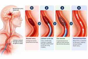
Illustration for educational purposes only. It is not intended to represent exact clinical anatomy or procedural outcomes. Clinical accuracy may vary.
Clinical trials have demonstrated improved functional outcomes with thrombectomy in appropriately selected patients with LVO. 2 Selected patients may benefit from treatment 6–24 hours after they were last known to be well. Eligibility depends on factors such as the location of the blockage, clinical condition and imaging findings. This extended window does not mean that every patient can benefit from late treatment. 1,3,4
Three Mechanical Thrombectomy Techniques, Explained Simply
1. Aspiration technique: A specialised catheter is advanced to the clot, and suction is applied to draw the clot into the catheter. Aspiration may be used alone or alongside other devices.
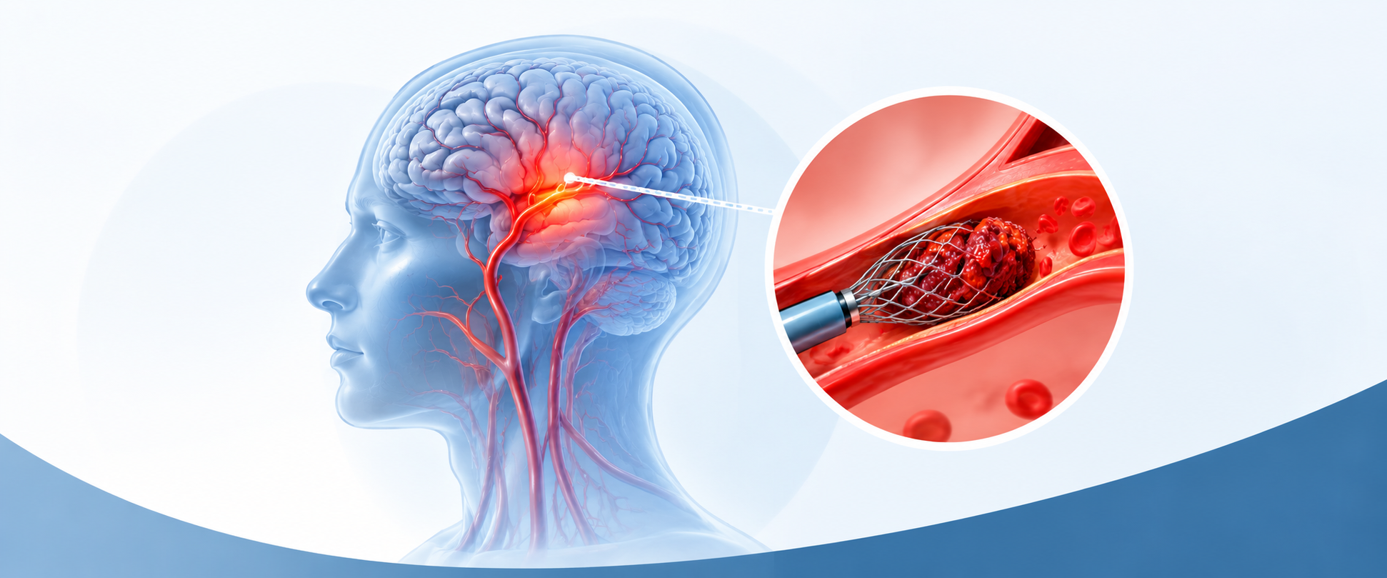
2. Stent retriever technique: A small, mesh-like device is passed through the clot and expanded to engage it. The device and captured clot are then carefully withdrawn through the catheter. Stent retrievers have played a central role in modern thrombectomy.
3. Combined Solumbra technique: The Solumbra approach combines a stent retriever with aspiration. The stent retriever engages the clot while an aspiration catheter provides suction near the blockage. This combines two clot-removal methods, although the choice of technique depends on the individual case and operator judgement.
Clinical trials have compared aspiration and stent-retriever approaches, supporting the use of different endovascular strategies in appropriate patients. 5,6 No single technique is best for every case.
How Has Mechanical Thrombectomy Changed Stroke Care?
The HERMES collaboration established strong evidence that endovascular thrombectomy improves functional outcomes in selected patients with LVO. 2 Subsequent trials, including DAWN and DEFUSE 3, demonstrated benefits for carefully selected patients treated in extended time windows. 3,4
These findings have changed stroke care by allowing clinicians to consider treatment beyond the earliest hours after symptom onset. Advanced imaging can help identify patients who may still benefit from restored blood flow. However, earlier treatment remains important, and recovery varies according to the extent of brain injury, successful reperfusion and subsequent care. 1
Why Does Every Minute Matter?
Brain tissue is highly sensitive to interrupted blood supply. Delays can allow irreversible injury to expand, reducing the likelihood of a good recovery. Rapid assessment, brain imaging, coordinated stroke teams and timely transfer to thrombectomy-capable centres are therefore essential. 1,2
Everyone should recognise stroke symptoms using B.E. F.A.S.T.: 7
- B — Balance: sudden loss of balance or coordination.
- E — Eyes: sudden visual changes.
- F — Face: facial drooping.
- A — Arms: weakness or numbness, particularly on one side.
- S — Speech: slurred speech or difficulty understanding words.
- T — Time: seek emergency medical help immediately.
A New Opportunity for Stroke Recovery
Mechanical thrombectomy has expanded treatment options for selected patients with acute ischaemic stroke. Its benefits depend on rapid recognition, appropriate patient selection and coordinated specialist care. Recognising stroke symptoms and seeking emergency treatment without delay can help patients reach the right care when every minute matters.
References
- Prabhakaran S, Gonzalez NR, Zachrison KS, et al. 2026 Guideline for the Early Management of Patients with Acute Ischemic Stroke: A Guideline from the American Heart Association/American Stroke Association. Stroke. 2026;57–e436. https://doi.org/10.1161/str.0000000000000513
- Goyal M, Menon BK, van Zwam WH, et al.; HERMES Collaborators. Endovascular thrombectomy after large-vessel ischaemic stroke: a meta-analysis of individual patient data from five randomised trials. Lancet. 2016; 387:1723–1731. https://doi.org/10.1016/s0140-6736(16)00163-x
- Nogueira RG, Jadhav AP, Haussen DC, et al.; DAWN Trial Investigators. Thrombectomy 6 to 24 hours after stroke onset or last known well. N Engl J Med. 2018; 378:11–21. 10.1056/NEJMoa1706442
- Albers GW, Marks MP, Kemp S, et al.; DEFUSE 3 Investigators. Thrombectomy for stroke at 6 to 16 hours with selection by perfusion imaging. N Engl J Med. 2018; 378:708–718. https://doi.org/10.1056/nejmoa1713973
- Lapergue B, Blanc R, Gory B, et al.; ASTER Trial Investigators. Effect of endovascular contact aspiration vs stent retriever on revascularization in patients with acute ischemic stroke and large vessel occlusion: the ASTER randomized clinical trial. JAMA. 2017; 318:443–452. https://doi.org/10.1001/jama.2017.9644
- Turk AS III, Siddiqui A, Fifi JT, et al.; COMPASS Investigators. Aspiration thrombectomy versus stent retriever thrombectomy as first-line approach for large vessel occlusion (COMPASS). Lancet. 2019; 393:998–1008. https://doi.org/10.1016/s0140-6736(19)30297-1
- American Stroke Association. Stroke Symptoms and Warning Signs. https://www.stroke.org/about-stroke/stroke-symptoms
Frequently Asked Questions
Yes. Selected patients may benefit up to 24 hours after symptom onset or last known well, depending on clinical assessment and imaging 1,3,4.
No. Thrombolysis uses medication to dissolve a clot, whereas thrombectomy uses catheter-based devices to remove it. Some eligible patients may receive both treatments 1.
No. It is primarily used for selected ischaemic strokes caused by accessible large-vessel blockages. It is not a treatment for every ischaemic stroke or for haemorrhagic stroke 1.
No. Restoring blood flow is an important treatment goal, but recovery also depends on the extent and location of brain injury, treatment timing, complications and rehabilitation.








Deje un comentario